Cognitive neuroscience
This article includes a list of general references, but it lacks sufficient corresponding inline citations. (December 2012) |
| Part of a series on |
| Psychology |
|---|
| Neuropsychology |
|---|
 |
Cognitive neuroscience is the scientific field that is concerned with the study of the biological processes and aspects that underlie cognition,[1] with a specific focus on the neural connections in the brain which are involved in mental processes.[2][3] It addresses the questions of how cognitive activities are affected or controlled by neural circuits in the brain. Cognitive neuroscience is a branch of both neuroscience and psychology, overlapping with disciplines such as behavioral neuroscience, cognitive psychology, physiological psychology and affective neuroscience.[4][2][3] Cognitive neuroscience relies upon theories in cognitive science coupled with evidence from neurobiology, and computational modeling.[2][3][4]
Parts of the brain play an important role in this field. Neurons play the most vital role, since the main point is to establish an understanding of cognition from a neural perspective, along with the different lobes of the cerebral cortex.
Methods employed in cognitive neuroscience include experimental procedures from psychophysics and cognitive psychology, functional neuroimaging, electrophysiology, cognitive genomics, and behavioral genetics.
Studies of patients with cognitive deficits due to brain lesions constitute an important aspect of cognitive neuroscience. The damages in lesioned brains provide a comparable starting point on regards to healthy and fully functioning brains. These damages change the neural circuits in the brain and cause it to malfunction during basic cognitive processes, such as memory or learning. People have learning disabilities and such damage, can be compared with how the healthy neural circuits are functioning, and possibly draw conclusions about the basis of the affected cognitive processes. Some examples of learning disabilities in the brain include places in Wernicke's area, the left side of the temporal lobe, and Broca's area close to the frontal lobe.[5]
Also, cognitive abilities based on brain development are studied and examined under the subfield of developmental cognitive neuroscience. This shows brain development over time, analyzing differences and concocting possible reasons for those differences.
Theoretical approaches include computational neuroscience and cognitive psychology.
Historical origins
[edit]
Cognitive neuroscience is an interdisciplinary area of study that has emerged from neuroscience and psychology.[6] There are several stages in these disciplines that have changed the way researchers approached their investigations and that led to the field becoming fully established.
Although the task of cognitive neuroscience is to describe the neural mechanisms associated with the mind, historically it has progressed by investigating how a certain area of the brain supports a given mental faculty. However, early efforts to subdivide the brain proved to be problematic. The phrenologist movement failed to supply a scientific basis for its theories and has since been rejected. The aggregate field view, meaning that all areas of the brain participated in all behavior,[7] was also rejected as a result of brain mapping, which began with Hitzig and Fritsch's experiments[8] and eventually developed through methods such as positron emission tomography (PET) and functional magnetic resonance imaging (fMRI).[9] Gestalt theory, neuropsychology, and the cognitive revolution were major turning points in the creation of cognitive neuroscience as a field, bringing together ideas and techniques that enabled researchers to make more links between behavior and its neural substrates.
While the Ancient Greeks Alcmaeon, Plato, Aristotle in the 5th and 4th centuries BC,[10] and then the Roman physician Galen in the 2nd century AD[11] already argued that the brain is the source of mental activity, scientific research into the connections between brain areas and cognitive functions began in the second half of the 19th century. The founding insights in the Cognitive neuroscience establishment were:
- In 1861, French neurologist Paul Broca discovered that a damaged area of the posterior inferior frontal gyrus (pars triangularis, BA45, also known as Broca's area) in patients caused an inability to speak.[12] His work "Localization of Speech in the Third Left Frontal Cultivation" in 1865 inspired others to study brain regions linking them to sensory and motor functions.[13]
- In 1870, German physicians Eduard Hitzig and Gustav Fritsch stimulated the cerebral cortex of a dog with electricity, causing different muscles to contract depending on the areas of the brain involved. This led to the suggestion that individual functions are localized to specific areas of the brain.[8]
- Italian neuroanatomist professor Camillo Golgi discovered in the 1870s that nerve cells could be colored using silver nitrate allowing Golgi to argue that all the nerve cells in the nervous system are a continuous, interconnected network.[14]
- In 1874, German neurologist and psychiatrist Carl Wernicke hypothesized an association between the left posterior section of the superior temporal gyrus and the reflexive mimicking of words and their syllables.[15]
- In 1878, Italian professor of pharmacology and physiology Angelo Mosso associated blood flow with brain functions. He invented the first neuroimaging technique, known as 'human circulation balance'. Angelo Mosso is a forerunner of more refined techniques like functional magnetic resonance imaging (fMRI) and positron emission tomography (PET).[16]
- In 1887, Spanish neuroanatomist professor Santiago Ramón y Cajal (1852–1934) improved the Golgi's method of visualizing nervous tissue under light microscopy by using a technique he termed "double impregnation". He discovered a number of facts about the organization of the nervous system: the nerve cell as an independent cell, insights into degeneration and regeneration, and ideas on brain plasticity.[17]
- In 1909, German anatomist Korbinian Brodmann published his original research on brain mapping in the monograph Vergleichende Lokalisationslehre der Großhirnrinde (Localisation in the cerebral cortex), defining 52 distinct regions of the cerebral cortex, known as Brodmann areas now, based on regional variations in structure. These Brodmann areas were associated with diverse functions including sensation, motor control, and cognition.[18]
- In 1924, German physiologist and psychiatrist Hans Berger (1873–1941) recorded the first human electroencephalogram EEG, discovering the electrical activity of the brain (called brain waves) and, in particular, the alpha wave rhythm, which is a type of brain wave.[19][20]
Origins in philosophy
[edit]Philosophers have always been interested in the mind: "the idea that explaining a phenomenon involves understanding the mechanism responsible for it has deep roots in the History of Philosophy from atomic theories in 5th century B.C. to its rebirth in the 17th and 18th century in the works of Galileo, Descartes, and Boyle. Among others, it's Descartes' idea that machines humans build could work as models of scientific explanation."[21] For example, Aristotle thought the brain was the body's cooling system and the capacity for intelligence was located in the heart. It has been suggested that the first person to believe otherwise was the Roman physician Galen in the second century AD, who declared that the brain was the source of mental activity,[22] although this has also been accredited to Alcmaeon.[23] However, Galen believed that personality and emotion were not generated by the brain, but rather by other organs. Andreas Vesalius, an anatomist and physician, was the first to believe that the brain and the nervous system are the center of the mind and emotion.[24] Psychology, a major contributing field to cognitive neuroscience, emerged from philosophical reasoning about the mind.[25]
19th century
[edit]Phrenology
[edit]
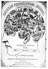
One of the predecessors to cognitive neuroscience was phrenology, a pseudoscientific approach that claimed that behavior could be determined by the shape of the scalp. In the early 19th century, Franz Joseph Gall and J. G. Spurzheim believed that the human brain was localized into approximately 35 different sections. In his book, The Anatomy and Physiology of the Nervous System in General, and of the Brain in Particular, Gall claimed that a larger bump in one of these areas meant that that area of the brain was used more frequently by that person. This theory gained significant public attention, leading to the publication of phrenology journals and the creation of phrenometers, which measured the bumps on a human subject's head. While phrenology remained a fixture at fairs and carnivals, it did not enjoy wide acceptance within the scientific community.[26] The major criticism of phrenology is that researchers were not able to test theories empirically.[6]
Localizationist view
[edit]The localizationist view was concerned with mental abilities being localized to specific areas of the brain rather than on what the characteristics of the abilities were and how to measure them.[6] Studies performed in Europe, such as those of John Hughlings Jackson, supported this view. Jackson studied patients with brain damage, particularly those with epilepsy. He discovered that the epileptic patients often made the same clonic and tonic movements of muscle during their seizures, leading Jackson to believe that they must be caused by activity in the same place in the brain every time. Jackson proposed that specific functions were localized to specific areas of the brain,[27] which was critical to future understanding of the brain lobes.
Aggregate field view
[edit]According to the aggregate field view, all areas of the brain participate in every mental function.[7]
Pierre Flourens, a French experimental psychologist, challenged the localizationist view by using animal experiments.[6] He discovered that removing the cerebellum (brain) in rabbits and pigeons affected their sense of muscular coordination, and that all cognitive functions were disrupted in pigeons when the cerebral hemispheres were removed. From this he concluded that the cerebral cortex, cerebellum, and brainstem functioned together as a whole.[28] His approach has been criticised on the basis that the tests were not sensitive enough to notice selective deficits had they been present.[6]
Emergence of neuropsychology
[edit]Perhaps the first serious attempts to localize mental functions to specific locations in the brain was by Broca and Wernicke. This was mostly achieved by studying the effects of injuries to different parts of the brain on psychological functions.[22] In 1861, French neurologist Paul Broca came across a man with a disability who was able to understand the language but unable to speak. The man could only produce the sound "tan". It was later discovered that the man had damage to an area of his left frontal lobe now known as Broca's area. Carl Wernicke, a German neurologist, found a patient who could speak fluently but non-sensibly. The patient had been the victim of a stroke, and could not understand spoken or written language. This patient had a lesion in the area where the left parietal and temporal lobes meet, now known as Wernicke's area. These cases, which suggested that lesions caused specific behavioral changes, strongly supported the localizationist view. Additionally, Aphasia is a learning disorder which was also discovered by Paul Broca. According to, Johns Hopkins School of Medicine, Aphasia is a language disorder caused by damage in a specific area of the brain that controls language expression and comprehension.[29] This can often lead to the person speaking words with no sense known as "word salad" [30]
Mapping the brain
[edit]In 1870, German physicians Eduard Hitzig and Gustav Fritsch published their findings of the behavior of animals. Hitzig and Fritsch ran an electric current through the cerebral cortex of a dog, causing different muscles to contract depending on which areas of the brain were electrically stimulated. This led to the proposition that individual functions are localized to specific areas of the brain rather than the cerebrum as a whole, as the aggregate field view suggests.[8] Brodmann was also an important figure in brain mapping; his experiments based on Franz Nissl's tissue staining techniques divided the brain into fifty-two areas.
20th century
[edit]Cognitive revolution
[edit]At the start of the 20th century, attitudes in America were characterized by pragmatism, which led to a preference for behaviorism as the primary approach in psychology. J.B. Watson was a key figure with his stimulus-response approach. By conducting experiments on animals he was aiming to be able to predict and control behavior. Behaviorism eventually failed because it could not provide realistic psychology of human action and thought – it focused primarily on stimulus-response associations at the expense of explaining phenomena like thought and imagination. This led to what is often termed as the "cognitive revolution".[31]
Neuron doctrine
[edit]In the early 20th century, Santiago Ramón y Cajal and Camillo Golgi began working on the structure of the neuron. Golgi developed a silver staining method that could entirely stain several cells in a particular area, leading him to believe that neurons were directly connected with each other in one cytoplasm. Cajal challenged this view after staining areas of the brain that had less myelin and discovering that neurons were discrete cells. Cajal also discovered that cells transmit electrical signals down the neuron in one direction only. Both Golgi and Cajal were awarded a Nobel Prize in Physiology or Medicine in 1906 for this work on the neuron doctrine.[32]
Mid-late 20th century
[edit]Several findings in the 20th century continued to advance the field, such as the discovery of ocular dominance columns, recording of single nerve cells in animals, and coordination of eye and head movements. Experimental psychology was also significant in the foundation of cognitive neuroscience. Some particularly important results were the demonstration that some tasks are accomplished via discrete processing stages, the study of attention,[33][34] and the notion that behavioural data do not provide enough information by themselves to explain mental processes. As a result, some experimental psychologists began to investigate neural bases of behaviour. Wilder Penfield created maps of primary sensory and motor areas of the brain by stimulating the cortices of patients during surgery. The work of Sperry and Gazzaniga on split brain patients in the 1950s was also instrumental in the progress of the field.[22] The term cognitive neuroscience itself was coined by Gazzaniga and cognitive psychologist George Armitage Miller while sharing a taxi in 1976.[35]
Brain mapping
[edit]New brain mapping technology, particularly fMRI and PET, allowed researchers to investigate experimental strategies of cognitive psychology by observing brain function. Although this is often thought of as a new method (most of the technology is relatively recent), the underlying principle goes back as far as 1878 when blood flow was first associated with brain function.[9] Angelo Mosso, an Italian psychologist of the 19th century, had monitored the pulsations of the adult brain through neurosurgically created bony defects in the skulls of patients. He noted that when the subjects engaged in tasks such as mathematical calculations the pulsations of the brain increased locally. Such observations led Mosso to conclude that blood flow of the brain followed function.[9]
Commonly the cerebrum is divided into 5 sections: the frontal lobe, occipital lobe, temporal lobes, parietal lobe, and the insula.[36] The brain is also divided into fissures and sulci.[37] The lateral sulcus called the Sylvian Fissure separates the frontal and temporal lobes. The insula is described as being deep to this lateral fissure. The longitudinal fissure separates the lobes of the brain length-wise. Lobes are considered to be distinct in their distribution of vessels.[36] The overall surface consists of sulci and gyri which are necessary to identify for neuroimaging purposes.[37]
Notable Experiments
[edit]Throughout the history of cognitive neuroscience, many notable experiments have been conducted. For example, the mental rotation experiment conducted by Kosslyn et al., 1993,[38] indicated that the time it takes to mentally rotate an object via imagination takes the same amount of time as actually rotating it; they found that mentally rotating an object activates parts of the brain involved in motor functioning, which may explain this similarity.[38]
Another experiment is describes the two mechanisms of processing visual attention: bottom-up attention, and top-down attention.[39] They define bottom-up attention is the brain visually processing salient images first, and then the surrounding information, while top-down attention involves focusing on task-relevant objects first. The researchers found that the ventral stream focuses on visual recognition, the dorsal stream is involved in the spatial information concerning the object.
As experiments in cognitive neuroscience, what these have in common is that the researchers are measuring activities or behaviors that we can see, and then determining the neural basis of the function and what part of the brain is involved.
Emergence of a new discipline
[edit]Birth of cognitive science
[edit]On September 11, 1956, a large-scale meeting of cognitivists took place at the Massachusetts Institute of Technology. George A. Miller presented his "The Magical Number Seven, Plus or Minus Two" paper[40] while Noam Chomsky and Newell & Simon presented their findings on computer science. Ulric Neisser commented on many of the findings at this meeting in his 1967 book Cognitive Psychology. The term "psychology" had been waning in the 1950s and 1960s, causing the field to be referred to as "cognitive science". Behaviorists such as Miller began to focus on the representation of language rather than general behavior. David Marr concluded that one should understand any cognitive process at three levels of analysis. These levels include computational, algorithmic/representational, and physical levels of analysis.[41]
Combining neuroscience and cognitive science
[edit]Before the 1980s, interaction between neuroscience and cognitive science was scarce.[42] Cognitive neuroscience began to integrate the newly laid theoretical ground in cognitive science, that emerged between the 1950s and 1960s, with approaches in experimental psychology, neuropsychology and neuroscience. (Neuroscience was not established as a unified discipline until 1971[43]). In the late 1970s, neuroscientist Michael S. Gazzaniga and cognitive psychologist George A. Miller were said to have first coined the term "cognitive neuroscience."[44] In the very late 20th century new technologies evolved that are now the mainstay of the methodology of cognitive neuroscience, including TMS (1985) and fMRI (1991). Earlier methods used in cognitive neuroscience include EEG (human EEG 1920) and MEG (1968). Occasionally cognitive neuroscientists utilize other brain imaging methods such as PET and SPECT. An upcoming technique in neuroscience is NIRS which uses light absorption to calculate changes in oxy- and deoxyhemoglobin in cortical areas. In some animals Single-unit recording can be used. Other methods include microneurography, facial EMG, and eye tracking. Integrative neuroscience attempts to consolidate data in databases, and form unified descriptive models from various fields and scales: biology, psychology, anatomy, and clinical practice.[45]

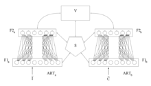
Adaptive resonance theory (ART) is a cognitive neuroscience theory developed by Gail Carpenter and Stephen Grossberg in the late 1970s on aspects of how the brain processes information. It describes a number of artificial neural network models which use supervised and unsupervised learning methods, and address problems such as pattern recognition and prediction.[46]
In 2014, Stanislas Dehaene, Giacomo Rizzolatti and Trevor Robbins, were awarded the Brain Prize "for their pioneering research on higher brain mechanisms underpinning such complex human functions as literacy, numeracy, motivated behaviour and social cognition, and for their efforts to understand cognitive and behavioural disorders".[47] Brenda Milner, Marcus Raichle and John O'Keefe received the Kavli Prize in Neuroscience "for the discovery of specialized brain networks for memory and cognition"[48] and O'Keefe shared the Nobel Prize in Physiology or Medicine in the same year with May-Britt Moser and Edvard Moser "for their discoveries of cells that constitute a positioning system in the brain".[49]
In 2017, Wolfram Schultz, Peter Dayan and Ray Dolan were awarded the Brain Prize "for their multidisciplinary analysis of brain mechanisms that link learning to reward, which has far-reaching implications for the understanding of human behaviour, including disorders of decision-making in conditions such as gambling, drug addiction, compulsive behaviour and schizophrenia".,[50]
Recent trends
[edit]Recently the focus of research had expanded from the localization of brain area(s) for specific functions in the adult brain using a single technology. Studies have been diverging in several different directions: exploring the interactions between different brain areas, using multiple technologies and approaches to understand brain functions, and using computational approaches.[51] Advances in non-invasive functional neuroimaging and associated data analysis methods have also made it possible to use highly naturalistic stimuli and tasks such as feature films depicting social interactions in cognitive neuroscience studies.[52]
In recent years, there have been a lot of new advancements in the field of Cognitive Neuroscience. One new technique that has emerged is called shadow imaging. This method has combined different aspects of various neuroimaging techinques to create one that is more versatile. It uses standard light microscopy and melds it with fluorescence labeling of the interstitial fluid in the brain's extracellular space. This technique can help researchers get a bigger and more detailed look at brain tissue. This can help researchers understand more on anatomy and viability for their experiments. This technique has helped to see neurons, microglia, tumor cells and blood capillaries more closely. Shadow imaging is a new approach that shows a lot of promise in the field of neuroimaging.[53]
Another very recent trend in cognitive neuroscience is the use of optogenetics to explore circuit function and its behavioral consequences.[54] This new technology is a combination of genetic targeting of certain neurons and using the imaging technology to see targets in living neurons. This technique allows scientists to see the neurons while they are still intact in animals and be able to trace the electrical happenings in that cell. This new technology has been used successfully in many experiments and it is helping researchers in observing brain activity and understanding its role in disease, behavior and function.[55]
Researchers have also modified a fMRI and made it more efficient, in a technique called direct imaging of neuronal activity or DIANA. This group of researchers changed the software to collect data every 5 milliseconds, which is 8 times faster than what the normal technique captures. After, the software can stitch together all of the images taken during the imaging and create a full slice of the brain.[56]
Cognitive Neuroscience and Artificial Intelligence
[edit]Cognitive neuroscience has played a major role in shaping artificial intelligence (AI). By studying how the human brain processes information, researchers have developed AI systems that simulate cognitive functions like learning, pattern recognition, and decision-making. A good example of this is neural networks, which are inspired by the connections between neurons in the brain. These networks form the foundation of many AI applications.[57]
Deep learning, a subfield of AI, uses neural networks to replicate processes similar to those in the human brain. For instance, convolutional neural networks (CNNs) are modeled after the visual system and have transformed tasks like image recognition and speech analysis. AI also benefits from advancements in brain imaging technologies, such as functional magnetic resonance imaging (fMRI) and electroencephalography (EEG). These tools provide valuable insights into neural activity, which help improve AI systems designed to mimic human thought processes.[58]
Despite the progress, replicating the complexity of human cognition remains a challenge. Researchers are now exploring hybrid models that combine neural networks with symbolic reasoning to better mimic how humans think and solve problems. This approach shows promise for addressing some of the limitations of current AI systems.[59]
Cognitive Neuroscience and Neurotherapy
[edit]Cognitive neuroscience contributed to development of novel noninvasive neurostimulation methods and developed in parallel with Neurotherapy aimed to address symptom control and cure several conditions in medical treatment.[60] Noninvasive neurotherapy have attracted significant attention from the scientific community since, these methods can be personalized and used in treatment independent of underlying conditions.[61] Based on research in cognitive neuroscience, Neurostimulation techniques apply different innovations to exert an energy-based impact on the nervous system by using electrical, magnetic, and/or electromagnetic energy to treat mental and physical health disorders in patients.[60][61] Since Neurotherapy implements systemic targeted delivery of an energy stimulus to a specific neurological zone in the body to alter neuronal activity and stimulate Neuroplasticity, the recent trend in the Cognitive neuroscience is the research of natural neurostimulation.[61]
Topics
[edit]- Attention
- Cognitive development
- Consciousness
- Creativity
- Decision-making
- Emotions
- Intelligence
- Language
- Learning
- Memory
- Perception
- Social cognition
- Mind Wandering
Methods
[edit]Experimental methods include:
- Psychophysics
- Eye-tracking
- Functional magnetic resonance imaging
- Electroencephalography
- Magnetoencephalography
- Electrocorticography
- Transcranial Magnetic Stimulation
- Computational Modeling
Notable people
[edit]- Jesper Mogensen, Danish neuroscientist and former university professor
See also
[edit]- Binding problem
- Cognitive biology
- Cognitive psychology
- Embodied cognition
- Experimental psychology
- Cognitive psychophysiology
- Affective neuroscience
- Social neuroscience
- Social cognitive neuroscience
- Cultural neuroscience
- List of cognitive neuroscientists
- Neurochemistry
- Neuroethology
- Neuroendocrinology
- Neuroscience
References
[edit]- ^ Gazzaniga, Ivry and Mangun 2002, cf. title
- ^ a b c Butler MJ, Senior C. "Toward an organizational cognitive neuroscience". Annals of the New York Academy of Sciences. 2007 Nov;1118(1):1-7. https://nyaspubs.onlinelibrary.wiley.com/doi/abs/10.1196/annals.1412.009
- ^ a b c Boone W, Piccinini G (2016). "The cognitive neuroscience revolution. Synthese". 2016 May;193:1509-34. https://link.springer.com/article/10.1007/s11229-015-0783-4
- ^ a b Gazzaniga 2002, p. xv
- ^ "Learning Disabilities | BRAIN". brainaacn.org. Retrieved 2022-04-27.
- ^ a b c d e Kosslyn, S, M. & Andersen, R, A. (1992). Frontiers in cognitive neuroscience. Cambridge, MA: MIT press.
- ^ a b Cordelia Erickson-Davis. "Neurofeedback Training for Parkinsonian Tremor and Bradykinesia" (PDF). Retrieved 2013-05-23.
- ^ a b c Fritsch, G.; Hitzig, E. (June 2009). "Electric excitability of the cerebrum (Über die elektrische Erregbarkeit des Grosshirns)". Epilepsy & Behavior. 15 (2): 123–130. doi:10.1016/j.yebeh.2009.03.001. PMID 19457461. S2CID 40594131.
- ^ a b c Raichle, Marcus E. (2009). "A brief history of human brain mapping". Trends in Neurosciences. 32 (2): 118–126. doi:10.1016/j.tins.2008.11.001. PMID 19110322. S2CID 205403489.
- ^ Guthri WKC (1971). A History of Greek Philosophy. London: Cambridge University Press. p. 348.
- ^ Lloyd G (2007). "Pneuma between body and soul". Journal of the Royal Anthropological Institute. 13: S135 – S146. doi:10.1111/j.1467-9655.2007.00409.x
- ^ Dronkers NF, Plaisant O, Iba-Zizen MT, Cabanis EA (May 2007). "Paul Broca's historic cases: high resolution MR imaging of the brains of Leborgne and Lelong". Brain. 130 (Pt 5): 1432–41. doi:10.1093/brain/awm042 https://academic.oup.com/brain/article-abstract/130/5/1432/283170?redirectedFrom=fulltext&login=false
- ^ Schiller F (1979). Paul Broca, Founder of French Anthropology, Explorer of the Brain. University of California Press. ISBN 978-0-520-03744-1., pp. 192–97
- ^ "Camillo Golgi – Facts". NobelPrize.org. Nobel Prize Outreach AB 2025. Tue. 14 Jan 2025. https://www.nobelprize.org/prizes/medicine/1906/golgi/facts/
- ^ Wernicke K (1995). "The aphasia symptom-complex: A psychological study on an anatomical basis (1875)". In Paul Eling (ed.). Reader in the History of Aphasia: From Franz Gall to Norman Geschwind. Vol. 4. Amsterdam: John Benjamins Pub Co. pp. 69–89. ISBN 978-90-272-1893-3.
- ^ Sandrone S (2014). "Weighing brain activity with the balance: Angelo Mosso's original manuscripts come to light". Brain. 137 (Pt 2): 621–633. doi:10.1093/brain/awt091 https://academic.oup.com/brain/article-abstract/137/2/621/280970?redirectedFrom=fulltext&login=false
- ^ Rozo JA, Martínez-Gallego I, Rodríguez-Moreno A (2024). "Cajal, the neuronal theory and the idea of brain plasticity". Front Neuroanat. 2024 Feb 19;18:1331666. doi: 10.3389/fnana.2024.1331666. https://pmc.ncbi.nlm.nih.gov/articles/PMC10910026/
- ^ Guillery RW (1999). "Brodmann’s ‘Localisation in the Cerebral Cortex’. Translated and edited by Laurence J. Garey". (pp. xviii+300; illustrated; £28 hardback; ISBN 1 86094 176 1.) London: Imperial College Press. 1999. Journal of Anatomy. 2000;196(3):493-496. doi:10.1046/j.1469-7580.2000.196304931.x
- ^ Haas LF (January 2003). "Hans Berger (1873–1941), Richard Caton (1842–1926), and electroencephalography". Journal of Neurology, Neurosurgery, and Psychiatry. 74 (1): 9. doi:10.1136/jnnp.74.1.9
- ^ İnce R, Adanır SS, Sevmez F (September 2021). "The inventor of electroencephalography (EEG): Hans Berger (1873-1941)". Child's Nervous System. 37 (9): 2723–2724. doi:10.1007/s00381-020-04564-z
- ^ Sirgiovanni, Elisabetta (2009). "The Mechanistic Approach to Psychiatric Classification" (PDF). Dialogues in Philosophy, Mental and Neuro Sciences. 2 (2): 45–49.
- ^ a b c Uttal, William R. (2011). Mind and Brain: A Critical Appraisal of Cognitive Neuroscience. MIT Press. ISBN 978-0-262-29803-2.[page needed]
- ^ Gross, Charles G. (July 1995). "Aristotle on the Brain". The Neuroscientist. 1 (4): 245–250. doi:10.1177/107385849500100408. S2CID 146717837.
- ^ Smith, C.U.M. (January 2013). "Cardiocentric Neurophysiology: The Persistence of a Delusion". Journal of the History of the Neurosciences. 22 (1): 6–13. doi:10.1080/0964704X.2011.650899. PMID 23323528. S2CID 34077852.
- ^ Hatfield, Gary (June 2002). "Psychology, Philosophy, and Cognitive Science: Reflections on the History and Philosophy of Experimental Psychology". Mind & Language. 17 (3): 207–232. doi:10.1111/1468-0017.00196.
- ^ Bear, Connors & Paradiso 2007, pp. 10–11.
- ^ Enersen, O. D. 2009
- ^ Boring, E.G. (1957). A history of experimental psychology. New York.
- ^ "Aphasia". www.hopkinsmedicine.org. Retrieved 2022-04-27.
- ^ "Wernicke area | Definition, Location, Function, & Facts | Britannica". www.britannica.com. Retrieved 2022-04-27.
- ^ Mandler, George (2002). "Origins of the cognitive (r)evolution". Journal of the History of the Behavioral Sciences. 38 (4): 339–353. doi:10.1002/jhbs.10066. PMID 12404267. S2CID 38146862.
- ^ "The Nobel Prize in Physiology or Medicine 1906".
- ^ Carrasco, Marisa (2011). "Visual attention: The past 25 years". Vision Research. 51 (13): 1484–1525. doi:10.1016/j.visres.2011.04.012. PMC 3390154. PMID 21549742.
- ^ Kastner, Sabine; Ungerleider, Leslie G. (2000). "Mechanisms of visual attention in the human cortex". Annual Review of Neuroscience. 23: 315–41. doi:10.1146/annurev.neuro.23.1.315. PMID 10845067. S2CID 11869810.
- ^ Gazzaniga, Michael (1984). "Preface". Handbook of Cognitive Neuroscience. pp. vii.
- ^ a b Casillo, Stephanie M.; Luy, Diego D.; Goldschmidt, Ezequiel (2020-02-01). "A History of the Lobes of the Brain". World Neurosurgery. 134: 353–360. doi:10.1016/j.wneu.2019.10.155. ISSN 1878-8750. PMID 31682988.
- ^ a b Ribas, Guilherme Carvalhal (February 2010). "The cerebral sulci and gyri". Neurosurgical Focus. 28 (2): E2. doi:10.3171/2009.11.FOCUS09245. ISSN 1092-0684. PMID 20121437.
- ^ a b Kosslyn, Stephen M.; Digirolamo, Gregory J.; Thompson, William L.; Alpert, Nathaniel M. (1998). "Mental rotation of objects versus hands: Neural mechanisms revealed by positron emission tomography". Psychophysiology. 35 (2): 151–161. doi:10.1111/1469-8986.3520151. ISSN 1469-8986. PMID 9529941.
- ^ Itti, L.; Koch, C. (March 2001). "Computational modelling of visual attention". Nature Reviews. Neuroscience. 2 (3): 194–203. doi:10.1038/35058500. ISSN 1471-003X. PMID 11256080.
- ^ Miller (1956). "The magical number seven plus or minus two: Some limits on our capacity for processing information". Psychological Review. 63 (2): 81–97. CiteSeerX 10.1.1.308.8071. doi:10.1037/h0043158. PMID 13310704. S2CID 15654531.
- ^ "Approaches in Cognitive Psychology". JungMinded.
- ^ not available, [1] not available
- ^ Society for Neuroscience. Date of the first meeting of the Society for Neuroscience
- ^ "About CNS". Cognitive Neuroscience Society. Retrieved 25 June 2023.
- ^ "Growth of Psychology as a Science - Origin of Psychology". www.boundless.com. Archived from the original on 28 June 2013. Retrieved 6 June 2022.
- ^ Carpenter, G.A., Grossberg, S., & Reynolds, J.H. (1991), ARTMAP: Supervised real-time learning and classification of nonstationary data by a self-organizing neural network Archived 2006-05-19 at the Wayback Machine, Neural Networks, 4, 565-588
- ^ "The Brain Prize". Archived from the original on 2015-09-05. Retrieved 2015-11-10.
- ^ "2014 Kavli Prize Laureates in Neuroscience". 2014-05-30.
- ^ "The Nobel Prize in Physiology or Medicine 2014". NobelPrize.org.
- ^ Gallager, James (6 March 2017). "Scientists win prize for brain research". BBC. Retrieved 6 March 2017.
- ^ Takeo, Watanabe. "Cognitive neuroscience Editorial overview" (PDF). Archived from the original (PDF) on 2012-12-24. Retrieved 2011-12-01.
- ^ Hasson, Uri; et al. (2004). "Intersubject Synchronization of Cortical Activity During Natural Vision". Science. 303 (5664): 1634–1640. Bibcode:2004Sci...303.1634H. doi:10.1126/science.1089506. PMID 15016991. S2CID 12688628.
- ^ Dembitskaya, Y., Boyce, A. K. J., Idziak, A., Pourkhalili Langeroudi, A., Arizono, M., Girard, J., Le Bourdellès, G., Ducros, M., Sato-Fitoussi, M., Ochoa de Amezaga, A., Oizel, K., Bancelin, S., Mercier, L., Pfeiffer, T., Thompson, R. J., Kim, S. K., Bikfalvi, A., & Nägerl, U. V. (2023). Shadow imaging for panoptical visualization of brain tissue in vivo. Nature communications, 14(1), 6411. https://doi.org/10.1038/s41467-023-42055-2
- ^ Pama, E. A. Claudia; Colzato, Lorenza S.; Hommel, Bernhard (6 September 2013). "Optogenetics as a neuromodulation tool in cognitive neuroscience". Frontiers in Psychology. 4: 610. doi:10.3389/fpsyg.2013.00610. PMC 3764402. PMID 24046763.
- ^ Deisseroth, Karl; Feng, Guoping; Majewska, Ania K.; Miesenböck, Gero; Ting, Alice; Schnitzer, Mark J. (2006-10-11). "Next-Generation Optical Technologies for Illuminating Genetically Targeted Brain Circuits". Journal of Neuroscience. 26 (41): 10380–10386. doi:10.1523/JNEUROSCI.3863-06.2006. ISSN 0270-6474. PMC 2820367. PMID 17035522.
- ^ Prillaman, McKenzie (2022-10-13). "Faster MRI scan captures brain activity in mice". Nature. doi:10.1038/d41586-022-03276-5. PMID 36229690.
- ^ LeCun, Yann; Bengio, Yoshua; Hinton, Geoffrey (2015-05-28). "Deep learning". Nature. 521 (7553): 436–444. Bibcode:2015Natur.521..436L. doi:10.1038/nature14539. ISSN 0028-0836. PMID 26017442.
- ^ Lake, Brenden M.; Ullman, Tomer D.; Tenenbaum, Joshua B.; Gershman, Samuel J. (2017). "Building machines that learn and think like people". Behavioral and Brain Sciences. 40: e253. arXiv:1604.00289. doi:10.1017/S0140525X16001837. ISSN 0140-525X. PMID 27881212.
- ^ Langley, Christelle; Cirstea, Bogdan Ionut; Cuzzolin, Fabio; Sahakian, Barbara J. (2022-04-05). "Theory of Mind and Preference Learning at the Interface of Cognitive Science, Neuroscience, and AI: A Review". Frontiers in Artificial Intelligence. 5. doi:10.3389/frai.2022.778852. ISSN 2624-8212. PMC 9038841. PMID 35493614.
- ^ a b Chapin TJ, Russell-Chapin LA (2013). "Neurotherapy and: Brain-based treatment for psychological and behavioral problems". Routledge; 2013 Dec 4.
- ^ a b c Val Danilov I (2023). "The Origin of Natural Neurostimulation: A Narrative Review of Noninvasive Brain Stimulation Techniques." OBM Neurobiology 2024; 8(4): 260; https://doi:10.21926/obm.neurobiol.2404260.
Further reading
[edit]- Baars, Bernard J.; Gage, Nicole M. (2010). Cognition, Brain, and Consciousness: Introduction to Cognitive Neuroscience. Academic Press. ISBN 978-0-12-381440-1.
- Bear, Mark F.; Connors, Barry W.; Paradiso, Michael A. (2007). Neuroscience. Lippincott Williams & Wilkins. ISBN 978-0-7817-6003-4.
- Churchland, Patricia Smith; Sejnowski, Terrence Joseph (1992). The Computational Brain. MIT Press. ISBN 978-0-262-33965-0.
- Code, Chris (2004). "Classic Cases: Ancient and Modern Milestones in the Development of Neuropsychological Science". In Code, Chris; Joanette, Yves; Lecours, André Roch; Wallesch, Claus-W (eds.). Classic Cases in Neuropsychology. pp. 17–25. doi:10.4324/9780203304112-8 (inactive 13 December 2024). ISBN 978-0-203-30411-2.
{{cite book}}: CS1 maint: DOI inactive as of December 2024 (link) - Enersen, O. D. (2009). John Hughlings Jackson. In: Who Named It. http://www.whonamedit.com/doctor.cfm/2766.html Retrieved 14 August 2009
- Gazzaniga, M. S., Ivry, R. B. & Mangun, G. R. (2002). Cognitive Neuroscience: The biology of the mind (2nd ed.). New York: W.W.Norton.
- Gallistel, R. (2009). "Memory and the Computational Brain: Why Cognitive Science will Transform Neuroscience." Wiley-Blackwell ISBN 978-1-4051-2287-0.
- Gazzaniga, M. S., The Cognitive Neurosciences III, (2004), The MIT Press, ISBN 0-262-07254-8
- Gazzaniga, M. S., Ed. (1999). Conversations in the Cognitive Neurosciences, The MIT Press, ISBN 0-262-57117-X.
- Sternberg, Eliezer J. Are You a Machine? The Brain, the Mind and What it Means to be Human. Amherst, NY: Prometheus Books.
- Ward, Jamie (2015). The Student's Guide to Cognitive Neuroscience (3rd ed.). Psychology Press. ISBN 978-1848722729.
- Handbook of Functional Neuroimaging of Cognition By Roberto Cabeza, Alan Kingstone
- Principles of neural science By Eric R. Kandel, James H. Schwartz, Thomas M. Jessell
- The Cognitive Neuroscience of Memory By Amanda Parker, Edward L. Wilding, Timothy J. Bussey
- Neuronal Theories of the Brain By Christof Koch, Joel L. Davis
- Cambridge Handbook of Thinking and Reasoning By Keith James Holyoak, Robert G. Morrison
- Handbook of Mathematical Cognition By Jamie I. D. Campbell
- Cognitive Psychology By Michael W. Eysenck, Mark T. Keane
- Development of Intelligence By Mike Anderson
- Development of Mental Processing By Andreas Demetriou, et al.
- Memory and Thinking By Robert H. Logie, K. J. Gilhooly
- Memory Capacity By Nelson Cowan
- Proceedings of the Nineteenth Annual Conference of the Cognitive Science
- Models of Working Memory By Akira Miyake, Priti Shah
- Memory and Thinking By Robert H. Logie, K. J. Gilhooly
- Variation in Working Memory By Andrew R. A. Conway, et al.
- Memory Capacity By Nelson Cowan
- Cognition and Intelligence By Robert J. Sternberg, Jean E. Pretz
- General Factor of Intelligence By Robert J. Sternberg, Elena Grigorenko
- Neurological Basis of Learning, Development and Discovery By Anton E. Lawson
- Memory and Human Cognition By John T. E. Richardson
- Society for Neuroscience. https://web.archive.org/web/20090805111859/http://www.sfn.org/index.cfm?pagename=about_SfN#timeline Retrieved 14 August 2009
- Keiji Tanaka,"Current Opinion in Neurobiology", (2007)
External links
[edit]- Cognitive Neuroscience Society Homepage
- There's Something about Zero
- What Is Cognitive Neuroscience?, Jamie Ward/Psychology Press
- goCognitive - Educational Tools for Cognitive Neuroscience (including video interviews)
- CogNet, The Brain and Cognitive Sciences Community Online, MIT
- Cognitive Neuroscience Arena, Psychology Press
- Cognitive Neuroscience and Philosophy, CUJCS, Spring 2002
- Whole Brain Atlas Top 100 Brain Structures
- Cognitive Neuroscience Discussion Group
- John Jonides, a big role in Cognitive Neurosciences by Beebrite
- Introduction to Cognitive Neuroscience
- AgliotiLAB - Social and Cognitive Neuroscience Laboratory founded in 2003 in Rome, Italy
Related Wikibooks


